
More photos of Melibe maugeana
May 25, 2006
From: Bill Rudman
To accompany Sabine Dittmann's message [#16693] about Melibe maugeana here are a few close-ups to show some features of the live animal. Species of Melibe lack a radula, and because their bodies are so thin they can lose much of their shape on preservation. They can also autotomise their cerata on preservation - or in the normal course of their life. Because of this, distinguishing species can be quite difficult. I suspect when we know more about this group, they should be identifiable from the shape and colour of the living animal. However, at present we don't have a good idea on variability within populations, so it is not yet possible to make good judgements on what constitutes infraspecific variation and what constitutes interspecific variation. Because of this a few more photos may be useful in the future.
Locality: Flinders Island, 6 m, [west coast of Eyre Peninsula], South Australia, Great Australian Bight, 21 June 2006, subtidal Posidonia seagrass bed. Length: 1 cm. Photographer: Sabine Dittmann.
Rudman, W.B., 2006 (May 25) More photos of Melibe maugeana. [Message in] Sea Slug Forum. Australian Museum, Sydney. Available from http://www.seaslugforum.net/find/16701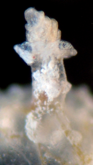
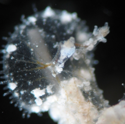
The upper two photos show dorsal and ventral views of the oral veil or oral hood, which is so effective as a net in which to catch small crustacea. In all the photos you will see traces of a fine translucent white or brownish network throughout the transparent body wall. This is sometimes described as a network of fibres or digestive gland ducts but it seems to be a mixture of muscle fibres, fine ducts of the digestive gland, and nerve fibres. If you look at the oral veil in the uppermost photo you can see 'fibres' radiating out from each rhinophore to the edge of the oral hood. These could be described as multiplex cables with nerve fibres to communicate with the sensory tentacles around the edge of the hood and muscle fibres to contract the hood when catching prey. The brown colouration shows that there is also digestive gland contents present as well. It is possible that like some other species of Melibe, this species contains symbiotic zooxanthellae - but I must emphasise that that is just a guess on my part.
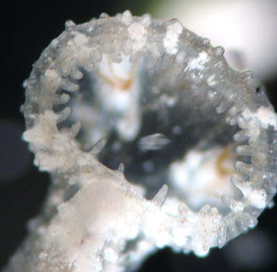
The second photo shows the oral hood from underneeth - a crustacean-eye view. Once the sensory tentacles make contact with a food-like item, the edge of the hood is contracted -as though it has a draw-string - and the potential prey trapped.
The bottom photo is of one rhinophore. In Melibe, the rhinophores are relatively small, perhaps a reflection of the diminished importance of chemoreception in Melibe in feeding, and the much greater importance of the sensory tentacles around the edge of the oral hood, .
Best wishes,
Bill Rudman
Related messages
-
Melibe maugeana from South Australia
From: Sabine Dittmann, May 25, 2006 -
Photos of Melibe maugeana from Melbourne
From: Harry Thorman, December 18, 2002 -
Melibe? from Port Philip Bay, Victoria
From: H. Thorman & A. McLaughlin, December 4, 2002 -
Melibe maugeana from nthn Tasmania
From: Bill Rudman, December 4, 2002