
Chromodoris naiki
- Anatomy
PHOTO
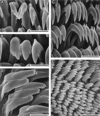
Chromodoris naiki, scanning electron micrographs. A. Inner lateral teeth, scale bar = 20 µm. B. Inner lateral teeth, scale bar = 15 µm. C. Lateral teeth from the central portion of the half-row, scale bar = 25 µm. C. Outer lateral teeth, scale bar = 25 µm. D. Jaw rodlets, scale bar = 20 µm. Radula by Ángel Valdés.
RELATED TOPIC
Chromodoris naiki is characterized internally by having the buccal mass divided evenly into an anterior glandular portion and a posterior muscular one. At the posterior end of the mass there are a pair of large, elongate salivary glands. The jaws are composed of a number of elongate, bifid rodlets about 15 µm in length. The radular formula is 38 x 41.0.41. Rachidian teeth are absent. The innermost lateral teeth have one large denticle on the inner side of the cusp and three to four denticles on the outer side. The remaining lateral teeth are hook-shaped, lack denticles on the inner side of the cusp and have a series of six to seven denticles along the outer edge. The outer laterals are elongate with six to seven denticles situated on the tips of the teeth.
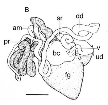
The reproductive system has an elongate and tubular ampulla that divides into the oviduct and the prostate. The oviduct is very short and enters the female glands near the center of the mass. The prostate is long, tightly coiled with several loops. It narrows into the muscular deferent duct. The deferent duct is also very long and coiled, and opens into a common atrium with the vagina. The penis is unarmed. The vagina is short and wide, slightly coiled. Near the end of the vagina the uterine duct emerges. It is short and convoluted and opens into the female glands. More proximally are the tightly coiled, digitiform seminal receptacle and the rounded, thin-walled bursa copulatrix.
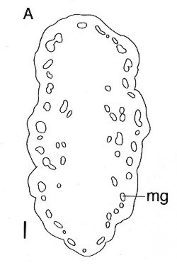
FIGURES: Chromodoris naiki. A. Disposition of the mantle glands, scale bar = 1 mm. B. General view of the reproductive system, scale bar = 1 mm. Abbreviations: am = ampulla, bc = bursa copulatrix, dd = deferent duct, fg = female glands, pr = prostate, sr = seminal receptacle, ud = uterine duct, v = vagina. Drawings: Ángel Valdés
Valdes, A., 2000 (July 23) Chromodoris naiki - Anatomy. [In] Sea Slug Forum. Australian Museum, Sydney. Available from http://www.seaslugforum.net/factsheet/chronaikanat