
Re: Chromodoris daphne feeding observation [2]
January 22, 2009
From: Leanne and David Atkinson
Concerning message #22154:
Dear Bill,
In answer to your question:
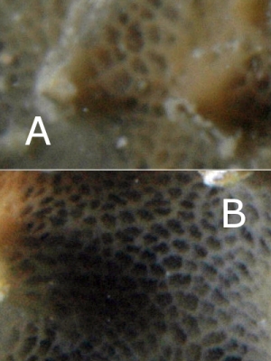
"I wondered if two sponge species were interwoven, but I suspect the bluish regions are paler parts of the Chelonaplysilla - perhaps where the slug has been feeding. I would be interested in your comments. Do you perhaps have a photo showing the join between blue and darker regions more clearly?"
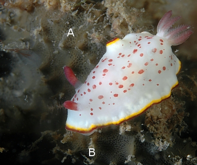
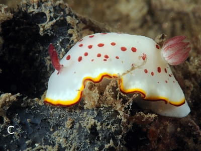
We have another photo of each nudibranch and sponge in the previous message. The two we sent in showed the mouth everted and indicated feeding better than the ones we didn't send. Here are the two other photos. We think what you are seeing is not two sponges but the hills and hollows of the sponge making shadows and light. We will keep looking for Chromodoris daphne feeding and see if we can come up with some photos that clear up the puzzle.
Locality: The Pipeline, Nelson Bay, Port Stephens and Fly Point, Port Stephens-Great Lakes Marine Park, Port Stephens , 5metres, New South Wales, Australia, Pacific Ocean, 09 January 2009 & 10 January 2009, Sandy silty bottom with sponges, ascidians, gorgonias, soft corals, hydroids and seaweed. Length: 30 mm. Photographer: Leanne and David Atkinson.
Regards,
Leanne and David Atkinson
atk@hunterlink.net.au
Thanks Leanne and David,
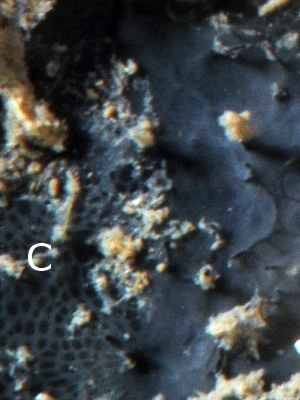
I think these extra photos have cleared up the puzzle quite well. Some sponges change colour in different parts of the same colony - certainly parts that are shaded are often much paler or almost colourlesss. It seems as though Chelonaplysilla violacea can range in colour from deep violet to dull blue to a reddish colour and also to a pale straw colour. One of the distinguishing features of Chelonaplysilla is its incorporation of small sand grains into its tissue which can usually be seen in C. violacea, in the granular appearance of the honeycomb-like surface of the colony. Interestingly your lower photo shows part of the honeycomb layer [C] being hidden by a smooth layer with raised conical papillae more typical of the related genera Darwinella. If I had seen the smooth section only I wouldn't have thought of it as Chelonaplysilla.
As I have often said, sponges are notoriously difficult to identify externally, and one of the reasons for this is that we don't have a good idea of the variability of living colonies. Although this is not a 'sponge forum' our growing collection of 'nudibranch food' photos is increasing our knowledge in both fields of biology.
Best wishes,
Bill Rudman
Related messages
-
Re: Chromodoris daphne from sthn Queensland
From: Denis Riek, January 23, 2009 -
Chromodoris daphne from sthn Queensland
From: Gary Cobb, January 20, 2009 -
Re: Chromodoris daphne feeding observation [2]
From: Leanne & David Atkinson, January 19, 2009 -
Chromodoris daphne feeding observation [2]
From: Bill Rudman, November 27, 2006 -
Chromodoris daphne feeding
From: Leanne & David Atkinson, November 27, 2006 -
Chromodoris daphne laying eggs
From: Leanne & David Atkinson, March 27, 2006 -
Octopus & Chromodoris daphne
From: Sean P. McMahon, November 29, 2002 -
Re: Chromodoris daphne
From: Wayne Ellis, January 18, 2001 -
Chromodoris daphne - where does it live?
From: Des Paroz, January 5, 2001