
Vayssierea felis - radular morphology
PHOTO
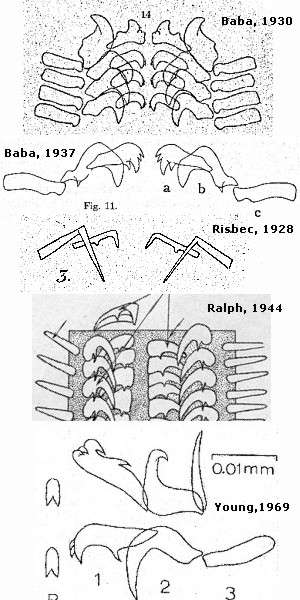
UPPER RIGHT: Drawings of the radula of Vayssierea felis under its various names, showing the different interpretations of its morphology.
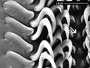
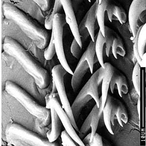
LOWER: Scanning Electron micrographs showing a half-row of the radula. Note remnant central tooth (arrowed). Presqu'île de Ouémo, Nouméa, New Caledonia, October 1988, on intertidal rocks with Spirorbis tubes. SEM PHOTO: Geoff Avern.
See Vayssierea felis page.
See Vayssierea felis References.
The shape of the radular teeth can be a very important character for taxonomists. In some cases it can tell us the family or genus an animal belongs to, and in some groups, when we are very fortunate, each species has distinct differences in the shape, or sometimes size of the teeth.
One important point is that the radular teeth are often very small, and before the development of scanning electron microscopes, a researcher's ability to accurately determine the shape of individual teeth was dependant on the quality of the light microscope available, the medium in which the radula was mounted for viewing, and the method used to extract and clean the tooth. One common problem was the use of Canada Balsam as a mounting medium. This has an almost identical refractive index as the protein of the radular teeth so when fine detail, such as denticles were looked at, strange optical tricks would occur which often meant that fine denticles would appear very coarse, and vice versa. Drawing radula from microscope mounts also led to many inaccuracies with badly aligned and adjusted camera lucida devices distorting dimensions.
The drawings I present here of the radula of Vayssierea felis well illustrate the difficulties we faced before the advent of Scanning Electron Microscopes.
This species has been found, under various names in many parts of the Indo-West Pacific. Vayssierea caledonica, from New Caledonia, Okadaia elegans from Japan and Pellibranchus cinnabareus from New Zealand, are all the same colour and the major features of their anatomy are identical. They all feed on spirorbid or similar tube worms, live in the intertidal zone and have direct development. The only feature distinguishing them has been the morphology of the radular teeth. In an animal so small, it is not surprising that different interpretations of the radular morphology have been reported from light microscope preparations. Risbec described a radula with two lateral teeth on each side and no rachidian, while both Ralph and Baba reported three lateral teeth on each side, the inner two teeth varying in denticulation in each species. More recently, Young has redescribed the teeth of specimens from Okinawa and reported for the first time a small rachidian tooth. I have prepared SEM mounts of specimens from New Caledonia which are identical to Young's description. I would consider all the earlier descriptions of the radular teeth to be attempts to describe the same morphology. Considering the reduced nature of the central tooth, it is possible that it may be absent in some populations or in some animals.
Rudman, W.B., 2000 (April 13) Vayssierea felis - radular morphology. [In] Sea Slug Forum. Australian Museum, Sydney. Available from http://www.seaslugforum.net/factsheet/radvaysfeli