Acanthodoris planca
Fahey & Valdés, 2005
Order: NUDIBRANCHIA
Suborder: DORIDINA
Superfamily: ANADORIDOIDEA
Family: Onchidorididae
DISTRIBUTION
Found only from Cove Rock, False Bay, South Africa.
PHOTO
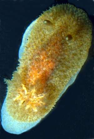
False Bay, South Africa. Photo: T.M.Gosliner
Etymology: Specific name planca is taken from the Greek word meaning Flat-footed, to describe the animal's prominent flat-footed appearance as it crawls.
The body shape of Acanthodoris planca is oblong and the foot extends beyond the mantle margin. The dorsum is covered with elongate papillae that appear short and conical when preserved. The papillae in the preserved specimens are longer at the mantle margin. The oral tentacles are short and blunt. The rhinophores are stout clubs that angle towards the posterior and have 15-17 lamellae. The gill is broad and spreads to cover the posterior third of the animal. There are ten gill leaves that are bi- and tripinnate. The background color of the dorsum is pale brown or orange and there is a darker orange patch of coloration mid-dorsum. The foot is white with tiny brownish-orange dots. The rhinophores and gill leaves are the same color as the background color, but the rhinophore tips are white.
Acanthodoris planca is externally most similar to the description of A. citrina Verrill, 1879, (a species that was later synonymized with A. pilosa by Thompson and Brown 1984) and to A. lutea MacFarland, 1925. All three species are orange yellow or orange and have a large gill that extends the width of the body. All three species have rhinophores that are similar in color as the body. The dorsum of each of these species is densely covered with conical tubercles. Acanthodoris planca has ten branchial leaves and both Verrill (1879) and MacFarland (1925) reported nine for both A. pilosa and A. lutea. The foot color differs between the species. Acanthodoris planca has a white foot with tiny brownish-orange spots, while that of A. lutea is orange yellow. Verrill did not report the foot color for A. citrina but the foot color of A. pilosa (Doris sparsa) was reported by Alder and Hancock (1846) as colorless and by Bergh (1879) as whitish or yellowish.
The radular morphology differs between these species. For example, Acanthodoris planca has densely arranged, multifid jaw rods, whereas A. lutea has triangular, hook-like structures. Verrill did not report the radular morphology of A. citrina but Thompson and Brown (1984) provided scanning electron micrographs of A. pilosa. Acanthodoris planca has 6-7 elongate, pointed outer lateral teeth, while A. lutea has 5-6 flattened, triangular plates "with a slight basal thickening" (MacFarland 1925). Acanthodoris pilosa has three outer lateral teeth that are flattened plates and a frail jaw cuticle with honeycombing (Thompson and Brown 1984).
There are reproductive differences among all three species as well.
-
Fahey, S.J. and Valdés, A. 2005. Review of Acanthodoris Gray, 1850 with a phylogenetic analysis of Onchidorididae Alder and Hancock, 1845 (Mollusca, Nudibranchia). Proceedings of the California Academy of Sciences 56(20): 213-272.
Fahey, S.J. & Valdés, A, 2005 (September 18) Acanthodoris planca Fahey & Valdés, 2005. [In] Sea Slug Forum. Australian Museum, Sydney. Available from http://www.seaslugforum.net/find/acanplan
Related messages
New Acanthodoris species from South Africa
September 19, 2005
From: Shireen Fahey
Greetings Bill,
I'd like to let you know about a new species of Acanthodoris that Angel Valdés and I have recently described. here is the Fact Sheet for Acanthodoris planca and images of the radula. The entire publication, which includes a phylogenetic analysis of the Onchidorididae can be downloaded from our website: www.calacademy.org/research/izg/nudibranchs/
Cheers,
Shireen
sfahey@calacademy.org
Fahey, S.J. and Valdés, A., 2005 (Sep 19) New Acanthodoris species from South Africa. [Message in] Sea Slug Forum. Australian Museum, Sydney. Available from http://www.seaslugforum.net/find/14780Radula of Acanthodoris planca
September 19, 2005
From: Shireen Fahey
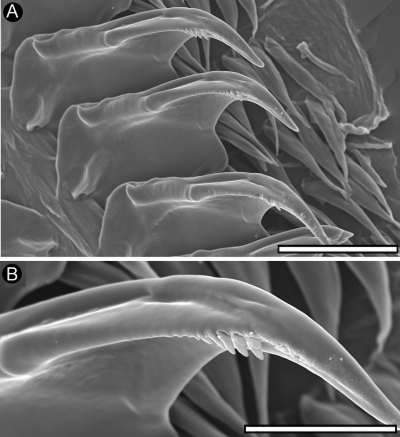
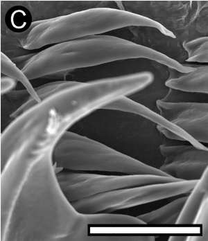
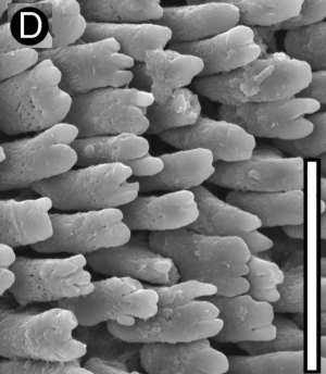
PHOTOS: Acanthodoris planca CASIZ 171754. Radular morphology. A First lateral teeth. Scale = 100 µm. B. Close up of denticles. Scale = 50 µm. C. Outer lateral teeth. Scale = 50 µm. D. Jaw rodlets. Scale = 30 µm. [Figure 20 - Fahey & Valdés, 2005]
These photos illustrate some of the features of the radular and jaw plate morphology of Acanthodoris planca as discussed on the Fact Sheet. It has densely arranged, multifid jaw rods, a few large denticles on the inner lateral radular tooth and 6-7 elongate, pointed outer lateral radular teeth.
-
Fahey, S.J. and Valdés, A. 2005. Review of Acanthodoris Gray, 1850 with a phylogenetic analysis of Onchidorididae Alder and Hancock, 1845 (Mollusca, Nudibranchia). Proceedings of the California Academy of Sciences 56(20): 213-272.
Shireen Fahey
billr@seaslugforum.net