
Aeolidiopsis ransoni
Pruvot-Fol, 1956
Order: NUDIBRANCHIA
Suborder: AEOLIDINA
Family: Aeolidiidae
PHOTO
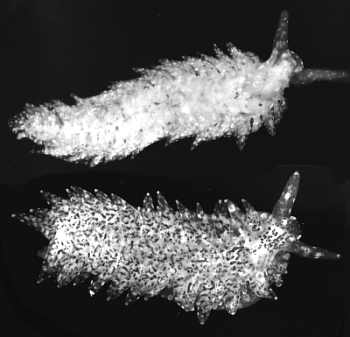
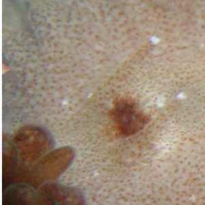
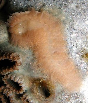
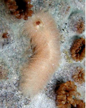
35 specimens, Lizard Island, Great Barrier Reef, Australia, off brown Palythoa growing in the shallow sub-littoral; June 4-10 1979. Specimens ranged in length from 8-16mm alive.
AM C124678-124688.
Upper: Black & White photo of white and brown speckled forms. Lower: colour photo of white form.
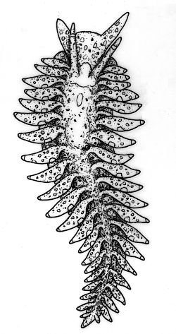
The body is elongate and flattened. The oral tentacles are broad and flattened, tapering to a rounded point. The rhinophores are smooth with a rounded tip. The cerata are flattened leaf-like structures and in smaller specimens (7 mm long) they are arranged in a single row along the edge of the dorsum, while in larger specimens (15 mm long) the cerata are more numerous. The cerata in front of the pericardium form a single horizontal row on each side, while the postpericardial cerata are arranged in closely packed, sloping rows of two to four cerata. In the living animals there is the impression of three horizontal rows of cerata, but in preserved animals and in dissection they are clearly short sloping rows. The cerata lie closely against the body forming a sinuous curve down the side and out over the foot. They point slightly posteriorly and are never held higher than the horizontal. The genital opening is on the right side below the third ceras from the front. The anus opens on the right, by a short papilla just above the cerata, behind the pericardium, either above and between the first and second or second and thrird post-pericardial rows. As mentioned earlier, the cerata are dorsoventrally flattened. The digestive gland in the ceras is branched, ramifying to all parts. The cnidosac is very prominent and contains large nematocysts from Palythoa. The foot is relatively broad, but does not extend out as far as the cerata. It is rounded anteriorly and does not extend into foot-corners.
There are two distinct colour forms. In one, the animal is translucent with small white patches all over. There is a distinct white patch between the rhinophores, and another midway along the posterior edge of each ceras. There is often a white band on the rhinophores just below the tip. The viscera show through as creamy white. There are a few golden brown specks scattered over the body, foot and cerata. When crawling over Palythoa, the translucency of the body renders it almost invisible. In the other colour form the animal appears speckled brown on the body, cerata, oral tentacles, rhinophores and foot. The brown colouration is from clusters of zooxanthellae in small branches of the digestive gland ramifying to all parts of the body.
See:
• A. ransoni - Biology and natural history.
• A. harrietae.
• Solar-powered Opisthobranchs
• Feeding on Palythoa
References:
• Rudman, W.B. (1982) The taxonomy and biology of further aeolidacean and arminacean nudibranch molluscs with symbiotic zooxanthellae. Zoological Journal of the Linnean Society, 74: 147-196.
Rudman, W.B., 2002 (July 18) Aeolidiopsis ransoni Pruvot-Fol, 1956. [In] Sea Slug Forum. Australian Museum, Sydney. Available from http://www.seaslugforum.net/find/aeolrans
Related messages
Rhinophores of Japanese Aeolidiopsis
July 25, 2002
From: Nishina Masayoshi
Dear Bill,
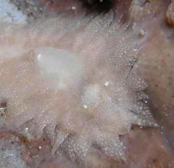
Thanks for the information about Aeolidiopsis. Concerning the shape of the rhinophores, here are some closeups of the heads of the brown speckled form. Sorry but I haven't a good photo showing the head in the white one. The upper right photo was taken on the 21 July 2002. The other two are from earlier photos.
Best Regards,
Nishina Masayoshi
nishina@wips.co.jp
Thanks Nishina,
The rhinophores are clearly smooth, which suggests, as I thought, that they are Aeolidiopsis ransoni.
Best wishes,
Bill Rudman
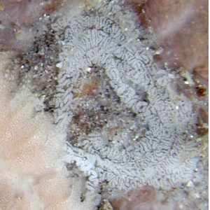
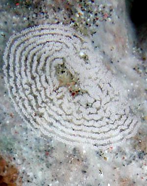
Egg ribbon of Japanese Aeolidiopsis
July 25, 2002
From: Nishina Masayoshi
Dear Bill,
Thanks for the information about Aeolidiopsis. Here is some more information on the egg masses. This photo was taken on the 21 July 2002 showing the brown speckled form laying an egg ribbon.
Best Regards,
Nishina Masayoshi
nishina@wips.co.jp
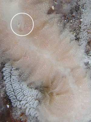
Dear Nishina,
Thanks for this photo. The egg ribbon in this photo is much more folded than in the earlier photo. This suggests to me that the variation you showed between the egg ribbons of the brown speckled animal and the white ones may just be part of the normal variation in the species, which would support my suggestion that they were one species.
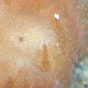
I have also included a close-up of the midregion of the animal because I think it shows (in ring) the anal papillae on the back just above the line of cerata. This is the position of the anus in A. ransoni, and combined with the smooth rhinophores shown in your other photos, makes me pretty convinced that this is A. ransoni.
Best wishes,
Bill Rudman
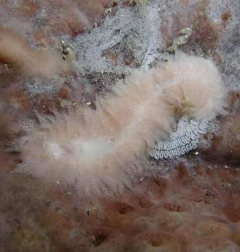
Aeolidiopsis ransoni from Hachijo Japan (1).
July 24, 2002
From: Nishina Masayoshi
Dear Bill,
These animals are a kind of Baeolidia such as Baeolidia palythoae Gosliner, 1985. I am not sure whether my animals are B. palythoae or not as I've not yet seen a photo of B. palythoae. I have found both brown speckled animals and white ones which I suspect are different species. I thought the whiter ones were perhaps a colour stage in animals that had not eaten for a while, but the shape of their egg mass looks different to the eggs of the brown spotted one. All of them eat and lay eggs on Palythoa tuberculosa and usually I can find 2 or 3 individuals on the same colony of Palythoa.
The photos here are of two brown speckled animals from the same colony of Palythoa. I have put photos of the white animals and its egg mass in a separate message.
Data: Hachijo Island, Japan, 9 June, 2002, Depth: 6m., Length: 20mm. on Palythoa tuberculosa. Photos: M. Nishina
Best Regards,
Nishina Masayoshi
nishina@wips.co.jp
Dear Nishina,
This is an interesting find. I think both the white and brown animals are the same species, Aeolidiopsis ransoni. Can you have a look at any other photos you have and see if you can see the rhinophores. In A. ransoni they should be smooth. To discuss your message, I gave just prepared some pages on this species and another Palythoa - feeder, Aeolidiopsis harrietae, which is very similar to Terry Gosliner's B. palythoae. In both those species the rhinophores have long papillae. I have discussed whether Aeolidiopsis or Baeolidia are the most appropriate genera for these species in a separate message.
But back to the white and brown animals. I have found this animal at both Lizard Island and Heron Island on the Great Barrier Reef, and it has two colour forms. One, like your animals is translucent with white speckling and a few brown spots, and the other is heavily speckled with dark brown. As you suspected the colour difference is 'food' related. The brown speckled animals retain zooxanthellae from the Palythoa, while the white ones do not. Whether the two colour forms are just phases in the digestive cycle of the slug, or represent distinct populations, one which harbours zooxanthellae and one that doesn't, I don't know. My observations suggested that the brown speckled form was found on the dorsal surface of the Palythoa while the white form was found burrowing just below the skin of the colony and could only be found by breaking off pieces of the colony and leaving them in a dish for a few hours until the white slugs crawled out. I must say that both the white and brown forms are very well camouflaged.
To the difference in the egg mass. It would certainly be worth getting some more information on this. You could be right and have two species here. However it is also possible that the egg ribbon in your separate message is aberrant. Egg ribbons are not always laid perfectly. It would be interesting to see if you can get some photos of more of these egg rbbons to see if they are consistently different. I have spread the information on A. harrietae and A. ransoni over a number of pages so jahve a look at the links on the Fact Sheet.
Best wishes,
Bill Rudman
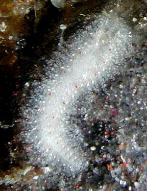
Aeolidiopsis ransoni from Hachijo Japan (2).
July 24, 2002
From: Nishina Masayoshi
Dear Bill,
To accompany my message about an aeolid feeding on Palythoa tuberculosa here are photos of the white colour form and its egg mass.
Hachijo Island, Japan, 7 July, 2002, Depth: 6m, Length: 20mm. Photos: M. Nishina
Best Regards,
Nishina Masayoshi
nishina@wips.co.jp
Masayoshi, N., 2002 (Jul 24) Aeolidiopsis ransoni from Hachijo Japan (2).. [Message in] Sea Slug Forum. Australian Museum, Sydney. Available from http://www.seaslugforum.net/find/7572Thanks Nishina,
As I said in your other message, I suspect this is the white colour form of Aeolidiopsis ransoni. I would need to know about the shape of the rhinophores to be sure.
Best wishes,
Bill Rudman.
Aeolidiopsis ransoni from Great Barrier Reef
July 24, 2002
From: Bill Rudman
Here are some black & white photos and a drawing of the Palythoa-feeding Aeolidiopsis ransoni from Lizard Island, Great Barrier Reef, Australia, off brown Palythoa growing in the shallow sub-littoral; June 4-10 1979. Specimens ranged in length from 8 to 16mm when alive. Further details on the biology of this fascinating Solar-powered nudibranch can be found on the Fact Sheet. Unfortunately I have no good colour photos of this species, although the black & white photos show the difference between the brown speckled form with zooxanthellae, and the white form, which lacks them. I have prepared a separate page with information on its biology and natural history.
Aeolidiopsis ranson was originally described by Pruvot-Fol (1956) from preserved specimens collected on Palythoa at Koukura Is, Tuamota Archipelago, in the eastern central Pacific. A number of inconsistencies between her description and my material are the result of Pruvot-Fol having only preserved material to study. She describes a peculiar animal in which the sole of the foot is rounded and replaces the sides of the body. This is not 'naturelle' as she surmises, but a result of preservation. Some of my preserved specimens, insufficiently narcotized, are similarly deformed. She describes the animals as having a reddish coloration which is also an artefact of preservation, many of my specimens becoming pinkish red in formalin. The absence of cnidosacs in Pruvot-Fol's specimens was also a result of preservation, as specimens can discharge all their nematocysts on preservation, thus rendering the cnidosac difficult to see. Pruvot-Fol erected the genus Aeolidiopsis for this species, citing the general body form, the single row of cerata, the dorsal position of the anus, and absence of cnidosacs, as features distinguishing it from other genera of the Aeolidiidae. Of these characters, the body form as described by her is an artefact of preservation. The single row of cerata along each side, found in smaller specimens, and the large number of almost horizontal rows of few cerata in larger specimens is unusual and is probably derived from the ceratal arrangement in Spurilla, described later in this paper. The fourth character, absence of cnidosacs, is, like the body shape, an error.
The reduced number of flattened cerata, arranged in almost lateral rows, is obviously an adaptation for symbiosis with zooxanthellae. Another distinctive feature is the single rounded anterior edge to the foot. The dorsal position of the anus and kidney opening is unique within the Aeolidiidae. While it is possible that the anal position is a primitive arrangement retained in this species, it is more probable that it is related to the unusual ceratal arrangement. The Aeolidiidae are in need of a generic revision which is outside the scope of this paper. Until this is undertaken I prefer to consider Aeolidiopsis a valid genus, but its relationship to other genera of the family is uncertain. I discuss this further in a separate message.
Best wishes,
Bill Rudman