
Aldisa banyulensis
Pruvot-Fol 1951
Order: NUDIBRANCHIA
Suborder: DORIDINA
Superfamily: EUDORIDOIDEA
Family: Dorididae
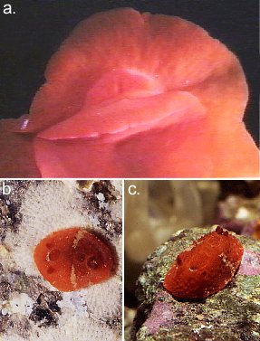
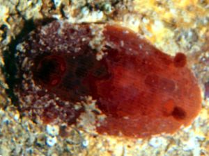
PHOTO
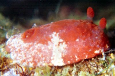
A, Ventral view of anterior end showing oral tentacles. B, 8 mm long, Punta del Vapor, Almuñecar, Granada, Spain. 6m deep, 20 October 1996. C, 18 mm long, Punta del Vapor, Almuñecar, Granada, Spain. 4m deep. 13 January, 2000. Photos: Luis Sanchez Tocino.
The identity of species of red species of Aldisa in the eastern Atlantic is somewhat confused. See A. smaragdina for a general discussion.
A. smaragdina, like A. banyulensis is red or orange-red with some white streaks on the mantle skirt. The mantle is pustulose and there are two darker pits in the dorsal midline between the gills and the rhinophores. The two differ in a number of external features. The rhinophores in A. smaragdina have few lamellae and they are arranged almost vertically while in A. banyulensis the more numerous lamellae are arranged horizontally. The gills in A. smaragdina are whitish in the upper half and red basally while in A. banyulensis they are a translucent red with no major white pigmentation. The rhinophore pockets in A. smaragdina have a series of tubercles around the edge and the gill pocket is strongly crenulate. In comparision, the edges of these pockets in A. banyulensis are smooth. Another more difficult difference to see is the shape of the head. In A. smaragdina the oral tentacles are slightly enrolled and bluntly cut off at the end, while in A. banyulensis they are triangular and flattened, with a pointed end. Both species have red eggs but they are larger [200µm diam] in A. banyulensis than in A. smaragdina [130µm].
Reference:
• Garcia, F.J., Garcia-Gomez, J.C. & Cervera, J.L. (1986) Ridescrizione di Aldisa banyulensis Pruvot-Fol, 1951 (Mollusca: Gastropoda: Nudibranchia). Lavori della Societe Italiana di Malacologia, 22: 97-110.
• Millen, S.V. & Gosliner, T.M. (1985) Four new species of dorid nudibranchs belonging to the genus Aldisa (Mollusca: Opisthobranchia), with a revision of the genus. Zoological Journal of the Linnean Society, 84: 195-233.
• Ortea, J.A., Perez, J.M. & Llera, E.M. (1982) Moluscos opistobranquios recolectados durante el plan de bentos circuncanario. Doridacea: primera parte (1). Cuadernos del Crinas, 3: 1-48.
• Pruvot-Fol, A. (1951) Etudes des nudibranches de la Mediterranee. 2. Arch. Zool. Exp. Gen., 88(1): 1-8O. (Pls. 14)
• Pruvot-Fol, A. (1953) Etude de quelques opisthobranches de la cote Atlantique du Maroc et du Senegal. Travaux de l'Institute Scientifique Cherifien, Zoologie, 5: 1-105. (Pls. 1-3)
Rudman, W.B., 2002 (November 11) Aldisa banyulensis Pruvot-Fol 1951. [In] Sea Slug Forum. Australian Museum, Sydney. Available from http://www.seaslugforum.net/find/aldibany
Related messages
Aldisa banyulensis - anatomy
November 18, 2002
From: Luis Sanchez Tocino
Dear Bill,
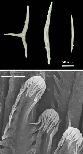
To accompany my earlier messages, here are some more photos of Aldisa banyulensis. These show the radula and the spicules in the mantle.
UPPER RIGHT: Upper photo shows 3 mantle spicules - Scale bar = 50µm. Lower shows the tip of the elongate radular teeth - Scale bar = 5µm
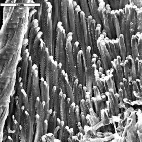
LOWER LEFT: SEM of section of radula showing teeth - Scale bar = 50µm
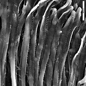
LOWER RIGHT: SEM of section of radula showing upper half of some teeth - Scale bar = 20µm
Luis
lstocino@ugr.es
Thanks Luis,
For those of you unfamiliar with the genus Aldisa, one of its most characteristic features is the shape of the radular teeth. Each transverse row can have up to 100 teeth in each half row, and the teeth are unusually thin and elongate with small denticles on the outside edge, near the tip. There are also a few large recurved denticles right at the tip of each tooth. Soem species can be recognised by the two pockets or pits found in the dorsal midline between the gills and the rhinophores, but this feature is not found in all species.
Best wishes,
Bill Rudman
Aldisa banyulensis from Spain (1)
November 13, 2002
From: Luis Sanchez Tocino
Dear Bill,
To accompany my messages on Aldisa smaragdina here is some comparative information on Aldisa banyulensis
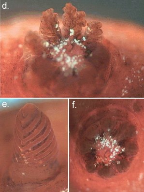
A, Ventral view of anterior end showing oral tentacles. B, 8 mm long, Punta del Vapor, Almuñecar, Granada, Spain. 6m deep, 20 October 1996. C, 18 mm long, Punta del Vapor, Almuñecar, Granada, Spain. 4m deep. 13 January, 2000. D, dorso-lateral view of gills. E, Rhinophore. F, dorsal view of gills. G, anterior end showing mantle tubercles. H, posterior end of mantle. I, SEM of mantle tubercles. Scale bar = 500µm.
To compare with Aldisa smaragdina here is some information on the number of gills and rhinophore lamellae in 3 specimens of different size.
8mm long - 9 gills - 8 rhino lamellae
10mm long - 9 gills - 9 rhino lamellae
18mm long - 8 gills - 9 rhino lamellae
Best wishes,
Luis
lstocino@ugr.es

Dear Luis,
Thanks for illustrating some of the external differences between these two species. Like species of Rostanga it is unfortunate that the differences are not usually clear in photographs of the whole animals.
Best wishes,
Bill Rudman
Aldisa banyulensis from Spain (2)
November 13, 2002
From: Luis Sanchez Tocino
Dear Bill,
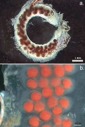
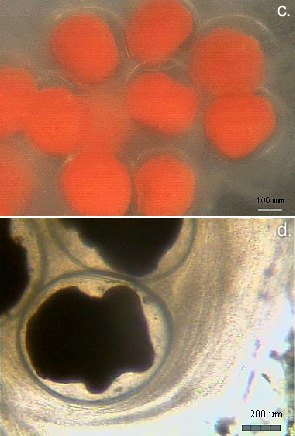
Here are some photos illustrating the egg ribbon and developing larvae in Aldisa banyulensis.
A, whole egg ribbon. Scale bar = 1mm. B-D, showing eggs and developing larvae. Scale bars: B = 200µm, C = 100µm, D = 200µm.
Luis
lstocino@ugr.es
Thanks Luis,
Bill Rudman
Aldisa smaragdina from Mediterranean Sea
November 1, 2001
From: Alma Sánchez
Dear Dr Rudman,
Here are two photos of Aldisa smaragdina Ortea, Perez & Llera, 1982. The specimen has been identified by Dr. Ángel Valdés.
This specimen was collected in August, 1999 at the Bay of Algeciras (Strait of Gibraltar, southern Spain) at 3 - 8 m. depth. Size: 15 – 20 mm. Place: Las Lajas Reef
Best wishes,
Alma Sánchez
almasanchez83@hotmail.com
Sánchez, A., 2001 (Nov 1) Aldisa smaragdina from Mediterranean Sea. [Message in] Sea Slug Forum. Australian Museum, Sydney. Available from http://www.seaslugforum.net/find/5589NOTE: See Luis Sanchez Tocino's message identifying this as A. banyulensis.
Thanks Alma,
Can anyone give some advice on identifying the Mediterranean species of Aldisa? I asked this question some time ago [see top of page] but have had no suggestions. For example how does A. smaragdina differ from Pruvot-Fol's A. binotata?
Best wishes,
Bill Rudman