
Re: Hypselodoris zebra larvae from Bermuda
February 20, 1999
From: J.E. Austin
Dear Dr. Rudman,
Thank-you for labelling my earlier pictures!! I can remember when I first looked at the larvae under the microscope, pulling this general invertebrate text open and still having no clue as what I was seeing. So exciting to have discovered something unknown!! Thanks to your prodding of course. I think there are great merits to any tinkering naturalist's eye for observation.
You mention that the veliger larvae have shells before they settle. If so, what are they made of? Are they like adult shells?
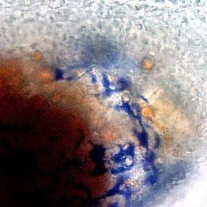
The larvae had no access to a food source while developing, so the blue color may come from stored food from the egg. I've attached a few more images, with some apparent internal structures I'm unable to make out and a little close view of a pigmented area.
PHOTOS: Upper Right: Blue pigmented streaks over ganglion.
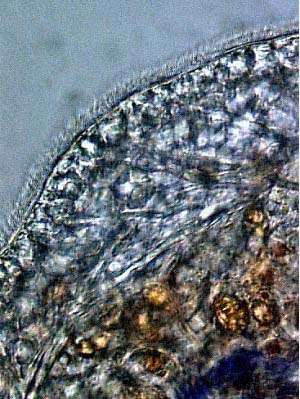
Lower Left: Cilia along edge of foot.
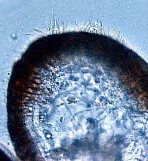
Lower Right: Large beating cilia around edge of velum.
Thank-you again for answering my questions.
Austin.
jaustin@bio.fsu.edu
Dear Austin,
Thanks again for the photos. First your question about larval shells. Gastropod larval shells have essentially the same makeup as adult shells, but the are normally smooth and unsculptured. There is an outer proteinaceous layer of a chitin-like substance called conchiolin and an inner layer of calcium carbonate. One very distinctive difference between opisthobranch larval shells and "prosobranch" larval shells is that in opisthobranchs the shell coils sinistrally not dextrally. To explain what that means - if you hold a snail shell, spire pointing up and the aperture facing you, the aperture will normally be on the right hand side (dextral coiling). In opisthobranch larvae the shell aperture is on the left hand side. This means that when the animal metamorphoses, the shell has to change its direction of coiling from sinistral to dextral. The larval shell (protoconch) sits very awkwardly on top of the adult shell and is described as "heterostrophic". All "heterobranch" gastropods (opisthobranchs, pulmonates, etc) have heterostrophic protoconchs.
One other distinction between opisthobranch and "prosobranch" larvae is that opisthobranchs only have one larval kidney rather than the pair found in "prosobranchs". Even though it is called a kidney I think its function is not really understood.
Cilia, like those you can see around the edge of the foot, also occur all over the sole of the foot, and are for locomotion. The much larger cilia on the velum, occur in a large band at the edge of the velar lobes and are for swimming. In feeding veligers there are also fields of cilia on the face of the velar lobes which direct feeding currents to the mouth.
I think the large golden-brown globules present in all the photos are lipid-filled globules, most probably the remnants of the food stores present in the yolk.
Best wishes,
Bill Rudman.
Related messages
-
Hypselodoris zebra - old reference and new photo
From: Thaddeus Murdoch, February 18, 2010 -
Photos of Hypselodoris zebra & eggs
From: Daniel Geiger, March 12, 1999 -
Re: Hypselodoris zebra larvae
From: Bill Rudman, February 26, 1999 -
Hypselodoris zebra larvae from Bermuda
From: J.E. Austin, February 18, 1999 -
re: Sea Slug Forum
From: J.E.Austin, November 11, 1998 -
Re: J.E.Austin's Hypselodoris zebra query
From: Bill Rudman, November 9, 1998