
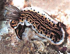
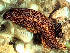
Hypselodoris zebra
(Heilprin, 1889)
Order: NUDIBRANCHIA
Suborder: DORIDINA
Family: Chromodorididae
DISTRIBUTION
Known only from Bermuda, tropical west Atlantic.
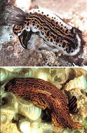
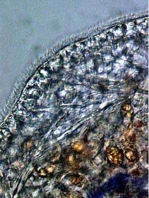
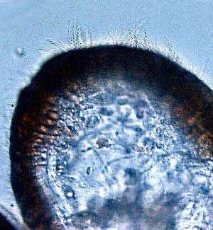
PHOTO
Bermuda, July 1991.
PHOTOS: Daniel Geiger. See message and photo of egg ribbon below.
RELATED TOPIC
Opisthobranch Defence - colour patterns
This chromodorid is not well-known and seldom illustrated, probably because its lives outside the "centres" of chromodorid diversity - the Indo-West Pacific, Caribbean, and Pacific east coast. Growing to over 18cm long it is one of the largest chromodorids. Hypselodoris zebra deserves an important place in "chromodorid history' because it was the species which attracted Crozier's attention and let to his early discussions and studies on the apparent immunity chromodorids have from fish predation.
References:
• Crozier,W.J. (1916). On the immunity coloration of some nudibranchs. Proceedings of the National Academy of Sciences, 2: 672-675.
• Crozier,W.J. & Arey, L.B. (1919). Sensory reactions of Chromodoris zebra. Journal of Experimental Zoology, 29: 261-310.
• Grode, S. H. & Cardellina, J.H., 1984. Sesquiterpenes from the sponge Dysidea etheria and the nudibranch Hypselodoris zebra. Journal of Natural Products, 47(1):76-83.
• Rudman, W.B. (1991). Purpose in pattern: the evolution of colour in chromodorid nudibranchs. Journal of Molluscan Studies, 57, (T.E. Thompson Memorial Issue):5-21.
See photos of larvae and egg ribbon below.
Authorship detailsRudman, W.B., 1999 (March 12) Hypselodoris zebra (Heilprin, 1889). [In] Sea Slug Forum. Australian Museum, Sydney. Available from http://www.seaslugforum.net/find/hypszebr
Related messages
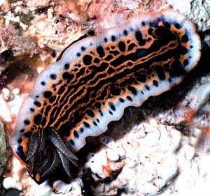
Hypselodoris zebra - old reference and new photo
February 18, 2010
From: Thaddeus Murdoch
Took a photo of a Hypselodoris zebra in Castle Harbour, Bermuda in January on a fringing reef in 2m depth. My research assistant was doing fish surveys concurrently and observed over 20 specimens within a 200-m by 10-m area. This highly clustered distribution perhaps confirms it's lecithotrophic life history strategy, as discussed in earlier posts for the species on your fine forum.
Below is a key reference for the species regarding it's endemic status in Bermuda which may be of use to your readers.
Locality: Castle Harbour, 2m, Bermuda, Atlantic Ocean, 28 January 2010, Fringing Reef within enclosed harbour. Length: 10 cm. Photographer: Thaddeus Murdoch.
- Thompson, T.E. (1977). The taxonomic status of two Bermudan Opisthobranchs. Journal of Molluscan Studies, 43: 218-222.
Thaddeus Murdoch
tjmurdoch@gov.bm
Murdoch T.J.T., 2010 (Feb 18) Hypselodoris zebra - old reference and new photo. [Message in] Sea Slug Forum. Australian Museum, Sydney. Available from http://www.seaslugforum.net/find/23230
Dear Thad,
Thanks for the photo. From other photos I have seen, it looks as though this species loses the white band around the edge of the mantle at it grows larger. Your colour form has many similarities to the west Atlantic form of H. picta.
I referred to Tom Thompson's paper in an earlier message [#622]. We can only hope that someone will consider the reproductive biology of the west Atlantic chromodorids to be worthy of study sometimes soon. Although some direct developers and lecithotrophic developers have restricted geographical distributions there are always examples which break the rules. As far as the local clustering you describe, I suspect this is in response to the presence of their preferred food sponge(s). Since veliger larvae settle and metamorphose on their food sponges, and the eggs of direct developers are usually laid on or near their food sponges, this sems to be the most likely cause of clustering, which is found in species of all development types.
Which brings me to the close-up I have included alongside. I am intersted in accumulating more data on the feeding specificity of the chromodorids so I always look carefully at photos for signs of potential food sponges. Most species of Hypselodoris feed on dysideid sponges. I am not sure if their is a ravaged dysideid colony in your photo or not. If you can see more in the original photo or have another photo which shows signs of such a sponge I would very much like a higher resolution scan to add to the Forum as a possibe feeding record.
Best wishes,
Bill Rudman
Photos of Hypselodoris zebra & eggs
March 12, 1999
From: Daniel Geiger
Dear Bill,
Here are some photos of Hypselodoris zebra which I took in Bermuda in July, 1991. I hope they are of some use. Thank you for the Haliotis pics.
Best wishes,
Daniel Geiger
Department of Biological Sciences
University of Southern California
Los Angeles,
CA 90089-0371
USA
http://nhm.org/~dgeiger/index.html
dgeiger@usc.edu
Dear Daniel,
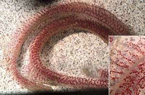
Thanks very much. The egg ribbon, looks like it was laid on an aquarium air stone, which I have found to be quite a favourite place for nudibranchs to lay their eggs. I have added an inset of a magnified section of the ribbon to show the spiral string of eggs within the egg coil. The relatively large eggs are typical of a species with non-feeding lecithotrophic larvae. I have also used the photos of the adults at the top of the page. It's nice to be able to put a picture with such an interesting animal.
Best wishes,
Bill Rudman.
Re: Hypselodoris zebra larvae
February 26, 1999
From: Bill Rudman
I have just noticed that Tom Thompson (1977) when discussing the limited distribution of Hypselodoris zebra says "it can be forecast that H.zebra will prove to have direct, non-pelagic development".
Austin's observations suggest that although its development is not direct, it is not very pelagic either, with non-feeding larvae quickly settling from the plankton.
Bill Rudman.
Reference: Thompson, T.E. (1977). The taxonomic status of two Bermudan Opisthobranchs. Journal of Molluscan Studies, 43: 218-222.
Rudman, W.B., 1999 (Feb 26). Comment on Re: Hypselodoris zebra larvae by Bill Rudman. [Message in] Sea Slug Forum. Australian Museum, Sydney. Available from http://www.seaslugforum.net/find/622Re: Hypselodoris zebra larvae from Bermuda
February 20, 1999
From: J.E. Austin
Dear Dr. Rudman,
Thank-you for labelling my earlier pictures!! I can remember when I first looked at the larvae under the microscope, pulling this general invertebrate text open and still having no clue as what I was seeing. So exciting to have discovered something unknown!! Thanks to your prodding of course. I think there are great merits to any tinkering naturalist's eye for observation.
You mention that the veliger larvae have shells before they settle. If so, what are they made of? Are they like adult shells?
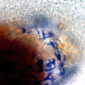
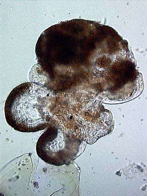
The larvae had no access to a food source while developing, so the blue color may come from stored food from the egg. I've attached a few more images, with some apparent internal structures I'm unable to make out and a little close view of a pigmented area.
PHOTOS: Upper Right: Blue pigmented streaks over ganglion.
Lower Left: Cilia along edge of foot.
Lower Right: Large beating cilia around edge of velum.
Thank-you again for answering my questions.
Austin.
jaustin@bio.fsu.edu
Dear Austin,
Thanks again for the photos. First your question about larval shells. Gastropod larval shells have essentially the same makeup as adult shells, but the are normally smooth and unsculptured. There is an outer proteinaceous layer of a chitin-like substance called conchiolin and an inner layer of calcium carbonate. One very distinctive difference between opisthobranch larval shells and "prosobranch" larval shells is that in opisthobranchs the shell coils sinistrally not dextrally. To explain what that means - if you hold a snail shell, spire pointing up and the aperture facing you, the aperture will normally be on the right hand side (dextral coiling). In opisthobranch larvae the shell aperture is on the left hand side. This means that when the animal metamorphoses, the shell has to change its direction of coiling from sinistral to dextral. The larval shell (protoconch) sits very awkwardly on top of the adult shell and is described as "heterostrophic". All "heterobranch" gastropods (opisthobranchs, pulmonates, etc) have heterostrophic protoconchs.
One other distinction between opisthobranch and "prosobranch" larvae is that opisthobranchs only have one larval kidney rather than the pair found in "prosobranchs". Even though it is called a kidney I think its function is not really understood.
Cilia, like those you can see around the edge of the foot, also occur all over the sole of the foot, and are for locomotion. The much larger cilia on the velum, occur in a large band at the edge of the velar lobes and are for swimming. In feeding veligers there are also fields of cilia on the face of the velar lobes which direct feeding currents to the mouth.
I think the large golden-brown globules present in all the photos are lipid-filled globules, most probably the remnants of the food stores present in the yolk.
Best wishes,
Bill Rudman.
Hypselodoris zebra larvae from Bermuda
February 18, 1999
From: J.E. Austin
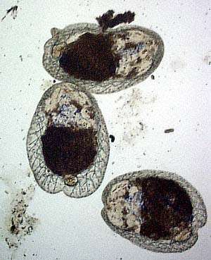
Last November, I wrote in concerning raising Hypselodoris zebra egg ribbons to hatching. I placed the egg ribbons in sea water (fresh from the reach off of the Bermuda Biological Station) changed daily. Ribbons were suspended in a cupped patch of plankton netting which was itself placed inside the cut open bottom of a 2 liter coke bottle turned upside down. At the mouth of the bottle was placed a cork with a tube inserted. The tube connected to an air pump. I changed the water daily by lifting up the plankton netting, transferring to a beaker and then switching the water. After maybe 3 wks., the eggs hatched into veliger larvae. They have cool patchs of purple presumably from internal food stores of the animals food source, the sponge Dysidea. Then the larvae became these little crawling beanie caps. Sorry for my inexpertise with sea slug life stages, but further description for any of my appended photos would be appreciated.
Sorry for taking so long to post these puppies. But the midnight when I discovered that they had hatched, I rounded up a poor graduate student to help me photograph them. Sea slugs are a driving inspiration.
J.E. Austin
Florida State University
jaustin@bio.fsu.edu
Dear Austin,
What great photos! It was certainly worthwhile you taking up my idea of seeing if you could get the eggs to develop. Your photos show that Hypselodoris zebra has lecithotrophic larvae. Lecithotrophic larvae are relatively short-lived non-feeding swimming veligers which metamorphose into crawling juveniles after a period in the plankton. In some cases the lecithotrophic veliger can spend only a few minutes or hours before settling, in other species they last for a few days before settling. Have you any idea how long the veligers spent swimming before settling?
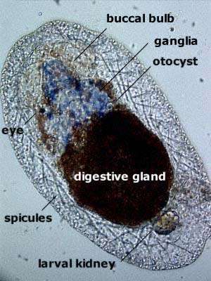
Your photos show (bottom left) a veliger which apparently has cast its shell and is about to settle down to benthic living. The black-edged lobes you can see are the reduced lobes of the velum - which with a band of beating cilia along the edge, are the veliger's swimming organ. In the photo (bottom right) of a metamorphosed juvenile, I have labelled some of the organs. I have never seen larvae with the brightly coloured pigmentation these ones do. The bluish purple region seems to include the buccal bulb and the nerve ganglia which cluster around the oesophagus just behind the buccal bulb and form its "brain".
The network of "spicules" form a structural network in the mantle tissue which is maintained in many dorid adults but is lost in most chromodorids. You have caught the larvae at such an early stage in their development that there does not seem to be any sign of rhinophore buds, and the larval kidney is still present. The otocysts are balance organs.
I am interested in the purple colouration. Did these animals have access to the sponge, Dysidea, on which the adults feed? If they did not, it suggests they carry pigment through from the egg - an interesting thought.
Thanks again Austin for sharing this interesting observation with us. It only goes to show that there are lots of valuable observations that can be made quite easily. One of the aims of the Forum is to publish such information. To me, and I presume to other researchers, such observations help to fill the many gaps in our knowledge. In themselves such observations usually are never published because they are too insignificant for a journal editor to bother with, or not "rigorous" enough for a scientific reviewer. To me they are all important pieces in a puzzle and I am happy to share them via the Forum.
Best wishes,
Bill Rudman.
re: Sea Slug Forum
November 11, 1998
From: J.E.Austin
Dear Dr. Rudman,
Thank-you so much for answering my sea slug questions. It began in Friday Harbor- the University of Washington's marine station. My passion for nudibranchs. I'm foremost a fish person, so I find that I'm straddling the vert-invert fence. More exciting that way. My name is Jenifer Austin but I simply use my last name, so I would appreciate if you just call me "Austin."
I will collect the Hypselodoris eggs today and work on raising them. There was a grad student form Tel Aviv raising Dorid eggs in a Larval ecology class up at Friday Harbor, so I'll see if I can find out his
regimes.
I am unaware of how collaborative information gets passed around the world of sea slug scientists. But if you are ever interested in Bermudean slugs, Wolfgang Sterrer's - Marine Flora and Fauna of Bermuda - provides an excellent guide with photos. Some collaborator from Florida collected nudibranchs here for him. I did not get to collect any Scyllea in the Sargassum because extratropical storm Mitch made an entrance with choppy wave action out on our deep water cruise.
I would like to ask your advice about research internships. I am planning to be in London for three months starting in May and am looking to do some research type internship preferable in a research lab at a University or Museum where the professor works on either sea slugs or fish. And has time to teach an eager student. I was wondering if you have any colleagues that you can recommend. As an American, I do not have much of a feel for the academic setting in the U.K. other than from websites and British students that I've met here in Bermuda.
Thank-you for your time,
Austin
Austin (J.E.)
Bermuda Biological Station for Research, Inc.
Ferry Reach
St. George, GE01
Bermuda
jaustin@bio.fsu.edu
jaustin@sargasso.bbsr.edu
xenomorph@micromyzon.com
jaustin@bio.fsu.edu
Austin, J.E., 1998 (Nov 11) re: Sea Slug Forum. [Message in] Sea Slug Forum. Australian Museum, Sydney. Available from http://www.seaslugforum.net/find/296Dear Austin,
Sorry to hear you were affected by "Mitch". I don't think I can really help with advice on possible research places in London. There are certainly no nudibranch workers there. John Taylor at the Natural History Museum might be able to give you advice.
Good luck with the egg raising and please keep us in mind if you find or photograph anything interesting.
Best wishes,
Bill Rudman
Re: J.E.Austin's Hypselodoris zebra query
November 9, 1998
From: Bill Rudman
I have split J.E.Austin's question(s) into separate topics .. Bill Rudman. This is on Hypselodoris zebra
Dear Dr. Rudman:
....... I have been watching Hypselodoris nudibranchs that were gathered outside the Bermuda Biological Station. They sit in my flow-through tank and have laid ribbons with red eggs. Some of them have little purple nodules beneath the side flaps of tissue. They are beautiful and hungry, I think. ......
You mention in Rudman, W.B. 1991, [Further studies on the taxonomy and biology of the octocoral-feeding genus Phyllodesmium, Ehrenberg, 1831 (Nudibranchia: Aeolidoidea). J. Moll. Stud. 57: 167-203], that Chromodorids can concentrate anti-feedants from consumed sponges. Has this been assayed in Hypselodoris zebra? This orange, blue-striped dorid found in Bermuda feeds on the purple sponge Dysidea etherea?. I have 6 specimens and would like to try something, any suggestions?
The paper makes reference to Rudman 1984, but I could not find the reference at the end and was wondering if you have that citation?
Thank-you for your time,
J.E. Austin
jaustin@bio.fsu.edu
I think the "little purple nodules beneath the side flaps of tissue" you describe are mantle glands, which are characteristic of chromodorids. Each genus has a different arrangement of these glands. Have a look at the photosof Chromodoris woodwardae and Mexichromis macropus.
As luck would have it I have just discovered a student review The Sequestering of Secondary Compounds from Sponges by Nudibranchs by Tim Judd, Colorado State University ,which has been recently posted. He refers to a fairly recent study (Grode, S. H., J. H. Cardellina II. 1984) on H. zebra and food sponge chemicals.
You ask what you can do with six specimens. I presume you mean while they are still alive? It seems they feed on the sponge Dysidea so food choice experiments would seem a bit of a waste of time. What we really need is some good behavioural experiments to show that fish find them distasteful, and associate the bad taste with particular colour patterns. What you need on hand is a good behavioural scientist who can help you set up a proper experimental regime.
Going from the sublime to the ridiculous. I am not sure whether Hypselodoris zebra has planktotrophic larvae, lecithotrophic larvae or direct development.
Perhaps if you have access to clean sea water you could keep some egg-masses alive and see whether the young hatch as veliger larvae or as crawling young. Without the proper equipment actually measuring egg and embryo diameter to get some idea of development type may be too difficult.
What would be nice, and this goes for many species is to get a better idea of the range of colour variation is found within a population. If you can find more specimens and a range of size, it would be useful to record the colour patterns, and which parts of the pattern seem to be constant in all specimens etc.
The last part of your question about Rudman 1984. That is the chromodorid generic review paper in which I describe the the mantle glands.
References: Grode, S. H., J. H. Cardellina II. 1984. Sesquiterpenes from the sponge Dysidea etheria and the nudibranch Hypselodoris zebra. Journal of Natural Products 47(1):76-83.
Rudman,W.B.,1984. The Chromodorididae (Opisthobranchia: Mollusca) of the Indo-West Pacific: a review of the genera. Zoological Journal of the Linnean Society, 81: 115-273.
Best wishes.... Bill Rudman
Rudman, W.B., 1998 (Nov 9). Comment on Re: J.E.Austin's Hypselodoris zebra query by Bill Rudman. [Message in] Sea Slug Forum. Australian Museum, Sydney. Available from http://www.seaslugforum.net/find/294