
Hallaxa michaeli - radula photos
July 24, 2006
From: Bill Rudman
I am posting today a Fact Sheet on Hallaxa michaeli. Here are some electron micrographs of the radular teeth of the specimen illustrated on that Fact Sheet.
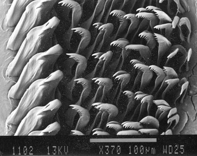
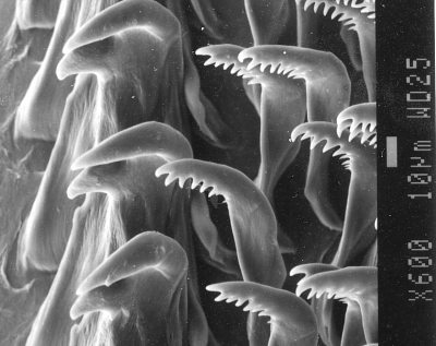
PHOTOS: Radular teeth and Jaw plates of H. michaeli. Barons Hut, Royal National Park, Sydney, March 1987, 20 m - on blue & white cunjevoi (ascidian), nudibranch 23 mm long. AM C152940. Upper photo: right side of radula showing 4 complete half rows. Scale bar = 100 µm. Middle photo: showing innermost tooth and inner laterals on right side. Scale bar = 10 µm. Lower photo: section of jawplate showing multi-tipped jaw rodlets.Scale bar = 10 µm. SEM Photos: Geoff Avern.
Rudman, W.B., 2006 (Jul 24) Hallaxa michaeli - radula photos. [Message in] Sea Slug Forum. Australian Museum, Sydney. Available from http://www.seaslugforum.net/find/17208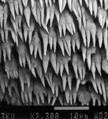
The family Actinocyclidae has interesting similarities in a number of anatomical features to the Chromodorididae and many workers consider the two families to have a common origin. One of the similarities is the morphology of the buccal armature, and if we exclude the innermost lateral tooth, both the radula and jaw plates, as illustrated here, could well belong to some species of the chromodorid genus Noumea.
Best wishes,
Bill Rudman
Related messages
-
Re: Hallaxa michaeli on food sponge
From: Leanne and David Atkinson, September 14, 2009 -
Re: Hallaxa michaeli on food sponge
From: Leanne and David Atkinson, August 31, 2009 -
Hallaxa michaeli on food sponge
From: Nicholas Missenden, March 23, 2007 -
Re: Hallaxa michaeli - feeding record
From: Nicholas Missenden, July 26, 2006 -
Hallaxa michaeli - feeding record
From: Nicholas Missenden, July 24, 2006