

Aeolidiopsis ransoni
Pruvot-Fol, 1956
Order: NUDIBRANCHIA
Suborder: AEOLIDINA
Family: Aeolidiidae
PHOTO
35 specimens, Lizard Island, Great Barrier Reef, Australia, off brown Palythoa growing in the shallow sub-littoral; June 4-10 1979. Specimens ranged in length from 8-16mm alive.
AM C124678-124688.
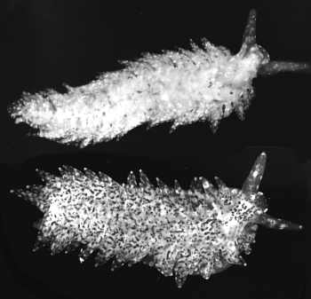
Upper: Black & White photo of white and brown speckled forms. Lower: colour photo of white form.
The body is elongate and flattened. The oral tentacles are broad and flattened, tapering to a rounded point. The rhinophores are smooth with a rounded tip. The cerata are flattened leaf-like structures and in smaller specimens (7 mm long) they are arranged in a single row along the edge of the dorsum, while in larger specimens (15 mm long) the cerata are more numerous. The cerata in front of the pericardium form a single horizontal row on each side, while the postpericardial cerata are arranged in closely packed, sloping rows of two to four cerata. In the living animals there is the impression of three horizontal rows of cerata, but in preserved animals and in dissection they are clearly short sloping rows. The cerata lie closely against the body forming a sinuous curve down the side and out over the foot. They point slightly posteriorly and are never held higher than the horizontal. The genital opening is on the right side below the third ceras from the front. The anus opens on the right, by a short papilla just above the cerata, behind the pericardium, either above and between the first and second or second and thrird post-pericardial rows. As mentioned earlier, the cerata are dorsoventrally flattened. The digestive gland in the ceras is branched, ramifying to all parts. The cnidosac is very prominent and contains large nematocysts from Palythoa. The foot is relatively broad, but does not extend out as far as the cerata. It is rounded anteriorly and does not extend into foot-corners.
There are two distinct colour forms. In one, the animal is translucent with small white patches all over. There is a distinct white patch between the rhinophores, and another midway along the posterior edge of each ceras. There is often a white band on the rhinophores just below the tip. The viscera show through as creamy white. There are a few golden brown specks scattered over the body, foot and cerata. When crawling over Palythoa, the translucency of the body renders it almost invisible. In the other colour form the animal appears speckled brown on the body, cerata, oral tentacles, rhinophores and foot. The brown colouration is from clusters of zooxanthellae in small branches of the digestive gland ramifying to all parts of the body.
See:
• A. ransoni - Biology and natural history.
• A. harrietae.
• Solar-powered Opisthobranchs
• Feeding on Palythoa
References:
• Rudman, W.B. (1982) The taxonomy and biology of further aeolidacean and arminacean nudibranch molluscs with symbiotic zooxanthellae. Zoological Journal of the Linnean Society, 74: 147-196.
Rudman, W.B., 2002 (July 18) Aeolidiopsis ransoni Pruvot-Fol, 1956. [In] Sea Slug Forum. Australian Museum, Sydney. Available from http://www.seaslugforum.net/factsheet/aeolrans
Related messages
-
Rhinophores of Japanese Aeolidiopsis
From: Nishina Masayoshi, July 25, 2002 -
Egg ribbon of Japanese Aeolidiopsis
From: Nishina Masayoshi, July 25, 2002 -
Aeolidiopsis ransoni from Hachijo Japan (1).
From: Nishina Masayoshi, July 24, 2002 -
Aeolidiopsis ransoni from Hachijo Japan (2).
From: Nishina Masayoshi, July 24, 2002 -
Aeolidiopsis ransoni from Great Barrier Reef
From: Bill Rudman, July 24, 2002